By Julia Schwarz
Ellen Zhong’s research creates detailed images of proteins and cells. But she isn’t using a microscope or a camera. She uses algorithms.

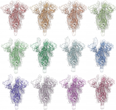
Zhong, an assistant professor of computer science, uses the tools of machine learning and computer vision to construct images from cryogenic electron microscopy (cryo-EM) experiments. Each cryo-EM experiment, she said, produces millions or even tens of millions of raw images. Her algorithms process this raw data to create clear, high-resolution images.
“The computational work,” Zhong said, “is crucial for extracting the signal from the noise.”
One example of this computational work is a method recently developed by Zhong and her collaborators, a technique called cryoFIRE. Their method can process image data exponentially faster than previous techniques, and without any loss of accuracy, lowering the computational burden required to create detailed images from each experiment. Advances like these, she said, make it easier, faster, and less expensive for researchers to process cryo-EM data.

Zhong’s own work primarily focuses on data related to protein structures, though the techniques she is developing can be applied to any type of cryo-EM experiments. Detailed images of proteins are critical, Zhong said, because understanding structure is essential to understanding function. Proteins are remarkably consistent in their shapes, so when they vary even slightly that variation can provide clues into their unique capability or function. Understanding protein structures has led to advances in antibody therapies and other medical applications.
The cryo-EM imaging field is also on the cusp of a very exciting leap right now, according to Zhong, because it’s recently become possible to “look directly inside cells,” instead of just at individual molecules like proteins or DNA. How individual molecules within a cell work together and interact is still largely a mystery, Zhong said, so getting a snapshot of what is happening inside a cell opens up exciting new possibilities for research across disciplines in bioengineering.
This article is from the Bioengineering: Unlocking mysteries, enabling impact issue of EQuad News magazine.